
Programmed Cell Death (Apoptosis)
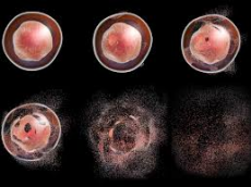
Programmed cell death (apoptosis) is the mechanism of removing unwanted, unnecessary, damaged or diseased cells from multicellular organisms.
Reasons for PCD:
Developmental importance
Numbers of cells need to be regulated and they fluctuate
Body function (sculpting body parts)
Diseased and damaged cells need to be removed
Necrosis is the death of cells, but it is not a programmed cell death. It has some distinctive hallmarks that separate it:
External trigger (unspecific)
No signal transduction
Inflammation
Cytoplasm swelling
Rupture of plasma membrane
Expansion of organelles
Hallmarks of apoptosis
Internal or external trigger
Signal transduction
Chromatin condensing
Nuclear fragmentation
Degradation in lysosomes
Cell shrinkage
There are two general cell death pathways: the intrinsic pathway and the extrinsic pathway.
How is apoptosis regulated?
The intrinsic pathway has a protein complex called the apotosome, and the extrinsic pathway has a complex called death inducing signalling complex (DISC), both work to activate caspases. Caspases are proteolytic enzymes used in apoptosis.
The extrinsic pathway begins with a death ligand (called fas ligand) from external origin binding to a death (fas) receptor on the plasma membrane of the target cell. This recruits a death domain, a death effector domain and procaspase 8 to the receptor site internally. This together forms the DISC complex. Procaspase 8 is cleaved to form active caspase 8. Caspase is an initiator caspase that activates effector caspases: caspase 3 and 7. These effector caspases carry out apoptosis.
Note: Death receptors are a part of the tumour necrosis receptor family.
Intrinsic pathway (apotosome formation)
The intrinsic pathway begins with an internal trigger activating the release of apaf1 (an adaptor protein). This causes apaf1 to go from locked conformation to semi-open autoinhibited conformation. Upon nucleotide exchange it forms a holoenzyme complex. Internal damage leads to changes in membrane outer membrane permeabilisation and mitochondrial transmembrane potential. BAX/BAK (part of the bcl-2 family) are reported to interact with and increase the opening of voltage gated anion channels, leading to change in membrane potential and release of cytochrome C. Calcium ions and ROS (reactive oxygen species) open the permeability transition pore complex in the inner membrane.
A quick recap:
Initiator caspase: Caspase 8 and Caspase 9
Effector caspase: Caspase 3 and Caspase 7
Regulators of apoptosis:
Bcl-2 family
Anti-apoptotic proteins
Inhibitors of apoptosis (IAP)
Anti-apoptotic proteins work by sequestering pro-apoptotic bcl-2 family members.
Inhibitors of apoptosis act as E3 ubiquitin ligases, ubiquitination caspases and causing their degradation.
Plants
Plants do carry out programmed cell death, but slightly differently. Plants do not have caspases nor do they have cytochrome c dependent apoptosis. Instead plants have metacaspases (a distant relative of caspases) and vacuolar processing enzymes.
Plant PCD plays a critical role in
Lignification (for plant support)
Organ development
Abiotic and biotic stress for example drought, heat, freezing, ozone, pathogens, UV light and nutrient depletion.
Biotrophic pathogens are pathogens that invade living plant cells. Necrotrophic pathogens are those that kill the cell before invading.
Biotrophic pathogens can be dealt with through hypersensitive immune reactions. Necrotrophic pathogens can be dealt with by transforming the plant cells to pocess the mammalian anti-apoptotic proteins.
image-https://www.google.com/url?sa=i&source=images&cd=&ved=2ahUKEwjM5onMi9DiAhXhyIUKHWg8DCoQjxx6BAgBEAI&url=http%3A%2F%2Fwww.biocompare.com%2FEditorial-Articles%2F332620-Monitoring-Apoptosis-by-Flow-Cytometry%2F&psig=AOvVaw2gOuV326hWdx14RW6jXdDv&ust=1559746752368893

0 Comment:
Be the first one to comment on this article.