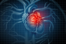

Myocardial infarction (Heart attack) and Angina and Breathing
Myocardial infarction causes of death:
Cardiac shock (decreased cardiac output)
Pulmonary edema (fluid in lungs)
Ventricular fibrillation
Occasionally heart rupture
Four factors that determine the likelihood of fibrillation
Injured tissue
Potassium depletion (hypokalaemia)
Muscle weakness
Sympathetic reflexes
Diagnosis
History of pain unrelated to exercise
ECG changes
Biochemical markers
Pain
Normally you cannot feel your heart beating, but there is a severe main caused by ischemia. Pain mediators are released (such as histamines) and this stimulates nerve endings.
ECG changes
With a lot of myocardial infarction comes ST segment elevation of the ECG trace (called STEMI). The cause of the elevation is unclear but it is a characteristic feature good for identifying. Within hours a patient will also develop an abnormal Q wave which will be present throughout life.
Biochemical Markers
Troponin isoform I (catalytic) is a protein found in cardiac muscle cells only. If we spot troponin I in the blood then it confirms muscle cell damage
Initial aims of treatment for Myocardial Infarction
Confirm diagnosis
Relieve pain
Stabilise hemodynamic abnormalities
Save as much myocardial tissue as possible
Treatment
Angioplasty (balloon tipped catheter that widens blood vessels and then a stent is used to keep them open)
Bypass surgery (redirect blood flow to restore blood flow)
Pharmacological drugs to prevent relapse
Recovery from MI occurs within a few months
Dead fibres enlarge and marginal fibres succumb, some non-functional muscle recovered. Fibrous tissue develops (structural tissue.) There is hypertrophy of normal areas to compensate.
Cardiac function after recovery
Pumping capacity permanently reduced
Cardiac reserve reduced
Diagnosis of angina
Angiography
Nuclear imaging
Stress test
Patient history
Changes in ECG during attack
Treatment for angina
GTN relieves pain
Vasodilators
Beta blockers block sympathetic enhancement of heart rate
Breathing
Breathing has three phases
Inspiration
Post-inspiration
Expiration
The phrenic nerve innervates the diaphragm. Breathing is controlled by a central pattern generator (we have talked about what these are previously) in the medulla oblongata (which is in the brain.) Therefore, damage to the medulla can have some serious consequences on breathing. The prebötzinger complex has a key role in generating a respiratory rhythm. Mechanisms of rhythm generation is still not fully understood, it could be networks, pacemakers or both. Substance P is a powerful modulator of the respiratory rhythm, if you apply it to the prebötzinger complex you get a big nerve burst with high frequencies. Substance P receptors are called NK1 receptors. If you attach a toxin (for example saporin) to substance P, when substance P binds to its receptor, it gets internalised and the toxin kills the cell. Severe unease in breathing is seen as a result.
The carotid sinus nerve in the carotid artery in the neck senses levels of oxygen. When levels are low, type I glomus cells release ATP which binds to receptors on the carotid sinus nerve, causing the nerves to fire to enhance breathing. If you knock out the receptors then the ability to sense low levels of oxygen is decreased. Therefore breathing is a plastic behaviour (adaptable.)
image-https://www.google.com/search?rlz=1C5CHFA_enGB759GB759&biw=1440&bih=721&tbm=isch&sa=1&ei=uYj2XPXAEIqqa97VooAF&q=heart+attack&oq=heart+attack&gs_l=img.3..0l10.151028.152563..152999...0.0..0.105.848.11j1......0....1..gws-wiz-img.......35i39j0i67.12exiMMPeF0#imgrc=Ptsfwnwtf-Aj1M:

0 Comment:
Be the first one to comment on this article.