
To study cells requires an understanding of how to use microscopes. Firstly we must distinguish the difference between magnification and resolution as these words are vital in your understanding on how big an organism is. Magnification is how much bigger the image is than the specimen. We can use a formula to calculate magnification.
The calculation is: Magnification = image size
___________
Object size
Resolution is how detailed an image is. For A level biology you especially need to know that the resolution is how well a microscope distinguishes between two points that are close together. This means that if you cannot see two separate objects are separate, then increasing the magnification will not make any difference in your understanding of the subject.
There are different types of microscopes, all with different resolutions and magnifications, they are as follows:
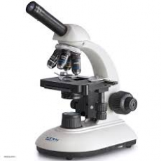
Light Microscopes
As you can guess by the name, these microscopes use light! The maximum resolution is about 0.2 micrometres so the smallest microscopic organisms will not be observed uses them. Usually, we will use light microscopes to look at whole cells or tissues. x1500 is the maximum useful magnification, a larger magnification will make the image appear blurry due to the limited resolution.
Laser Scanning Confocal Microscopes (a special type of light microscope)
Laser Scanning Confocal Microscopes produce a much clearer image than normal light microscopes. Firstly a specimen is usually tagged with a fluorescent dye. These lenses use laser beams which are intense beams of light to scan the specimen. The light beams cause the dye to give off light (fluoresce) and then the light is focused onto a detector through a pinhole. Meanwhile, the detector is connected to a computer and then an image is produced. The image can be 2-dimensional or 3-dimensional. The purpose of the pinhole is so that any out-of-focus light is blocked. One of the main advantages of these microscopes is that they can be used to look at objects at different depths in thick specimens.
Electron Microscopes
There are two types of electron microscopes; transmission and scanning. They both use electrons rather than light. The advantage of using these microscopes is that they have higher resolutions and thus give more detailed images, and smaller specimens can be observed. These microscopes are highly advanced, and so require specialist workers. TEM’s give a higher resolution and magnification than SEM’s.
Transmission electron microscope (TEM)
TEM’s spread a beam of electrons that are focused using electromagnets, these electrons pass through a specimen. Unlike light microscopes, the specimen is dead as the technique cannot work on living specimens. The more dense the specimen, the more electrons that are absorbed. Dense areas on a specimen produce a darker image thus there are variations of colour produced in an image, however the image produced is black and white. These microscopes are useful due to the high resolutions, and we can use them to observe a wide range of organelles. However the disadvantage to them is that they are expensive, and can only be used on thin specimens.
Scanning electron microscope (SEM)
These work by scanning a beam of electrons across a specimen, these electrons then knock of electrons from the specimen. The knocked off electrons are gathered in a cathode ray tube to form an image. The produced image is 3-dimensional, making SEM’s highly useful.
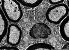
Image credits:
https://profilab24.com/media/image/product/25533/md/kern-transmitted-light-microscope-obe-1.jpg
https://www.news-medical.net/image.axd?picture=TEM%20schwann%20cell%20-%20Jose%20Luis%20Calvo%20_thumb%5B8%5D_thumb.jpg

0 Comment:
Be the first one to comment on this article.