
Absorbance and Spectrophotometry Assay
Differently coloured biochemical substances absorb visible light at varying wavelengths. Using a spectrophotometer, you can use the absorbance spectrum to determine the concentration of a solution. Measurements of the absorbance spectrum can be highly useful in identifying compounds or estimating the purity of them. Using different wavelengths, you can measure the absorbance of a known solution and can then compare the results to those of an unknown solution. We can thus explore similarities between compounds and work out what an unknown solution contains or its concentration.
Analytical methods require calibration, we use spectrophotometry to prepare a calibration curve for our solutions. The design of the spectrophotometry experiment was to carry out a series of tests on different solutions (methylene blue, carmine red and an unknown solution) and use the results for comparison. Once the maximum wavelength for peak absorption is established, we can determine the concentration of an unknown solution of methylene blue. We can then use Beer’s Law which demonstrates that the absorbance is proportional to the solution’s molar absorptivity and the concentration of a solute.
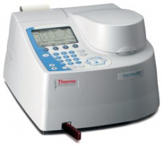
To begin the experiment, we firstly need to set the spectrophotometer and allow time for calibration using water placed in a cuvette. To do so, we change the wavelength to 350nm and then set the machine to blank. The water is therefore our control group. We prepare our samples, placing the same volume of methylene blue, carmine red and unknown solution in separate cuvettes (1mL), thus a second variable that was controlled. Our independent variable is wavelength ranging from 350nm- 700nm with predominantly 25nm intervals (with the exception of recordings taken around the maximum absorption peak.) Between each wavelength recording, we would blank with water, and plot the results on separate graphs for each sample. The absorbance values are our dependent values.
Next, we need to do a dilution experiment using methylene blue with the same wavelength for each dilution. The wavelength should be 695nm as this is the highest absorption peak. We can use concentrations from 0.0-5.0 at 1.0 intervals and use water to calibrate the spectrophotometer between each test.
With our collected results, we should complete a scatter graph. Open up excel, plot your data then produce a scientific graph. It will need legends, axis titles and a brief description of what the graph is demonstrating. Next, we need to explain what we can see, what standard curve graphs are produced. After we have described what we can see, we can think about why we see it. This is where you have to use your results, and think about it in a biological context. Perhaps comparing our results with published results. Are they similar or dissimilar; why? Then perhaps think about how it could have been improved. Did you answer the original question, have you managed to complete the aim of the experiment? It is useful to site references, as this will strengthen what you are trying to display. Perhaps do the experiment again, is it repeatable?
Image Credits: https://assets.thermofisher.com/TFS-Assets/CMD/product-images/F81552~p.eps-250.jpg

0 Comment:
Be the first one to comment on this article.